The Human Brain. Illustration Showing The Different Areas Of The Brain's Left Hemisphere Left And Right Hemisphere Above Right. See Image No. 11869...
digital illustration of how human brain uses blindsight to visualize moving tennis ball - 丘腦 幅插畫檔、美工圖案、卡通及圖標
Illustration of the location of the black substance, blue). This area contains dopamine neurons that are produced in smaller quantities in the case...前額葉皮質, 大腦皮質, 插圖
coronal view of a human brain in parkinsons disease. yellow surface is the thalamus and blue/green areas highlight fibers of the motor system - 丘腦 個照片及圖片檔磁力共振掃描
Brain, Drawing, Furrows That Determine The Lobes, They Bear The Names Of The Bones That Cover Them, Frontal, Parietal, Temporal, Occipital, Here It...
The Brain Is Constituted Of 2 Hemispheres Linked By Nerve Fibers Corpus Callosum. With The Brain Stem And The Cerebellum Joined In Back Derivation...大腦皮質
Cerebrovascular Disease Mri 1/2Recent Cerebrovascular Accident Of The Left Thalamus. Colorized Mri. Lesion Is Shown Here In Pink.
hippocampal sulcus remnant cysts seen on t2 axial mri (magnetic resonance imaging) - 丘腦 個照片及圖片檔磁力共振掃描
gray & white matter; spinal cord, 25x at 35mm. shows: gray matter (neuron cell bodies and unmyelinated nerve fibers), and white matter (myelinated nerve fibers in cross section). - 丘腦 個照片及圖片檔
digital illustration of unconscious and conscious pathways in human brain passing through thalamus ending in parietal lobe of cortex - 丘腦 幅插畫檔、美工圖案、卡通及圖標插圖
digital illustration of pain signal to area of somatosensory cortex in human brain via spine - 丘腦 幅插畫檔、美工圖案、卡通及圖標
digital illustration of head in profile showing direction of activating signals to cerebral cortex received from reticular activating system via thalamus - 丘腦 幅插畫檔、美工圖案、卡通及圖標
digital illustration of thalamus, hippocampus, and reticular formation highlighted in human brain - 丘腦 幅插畫檔、美工圖案、卡通及圖標人體構造
digital illustration of head in profile showing thalamus (grey), and frontal lobe (green) in brain - 丘腦 幅插畫檔、美工圖案、卡通及圖標
digital illustration of head in profile showing areas of brain used for processing emotion - 丘腦 幅插畫檔、美工圖案、卡通及圖標
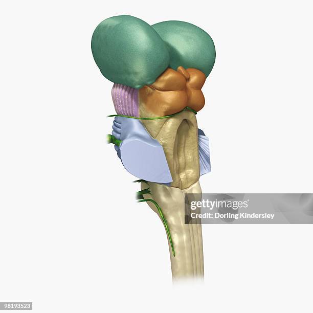
digital illustration of human brain stem with cerebellum removed revealing medulla and axons - 丘腦 幅插畫檔、美工圖案、卡通及圖標
digital illustration of head in profile showing brain highlighting thalamus in green and hypothalamus and pituitary gland in blue - 丘腦 幅插畫檔、美工圖案、卡通及圖標白色的背景
digital illustration of head in profile showing separate areas of brain highlighted in different colours - 丘腦 幅插畫檔、美工圖案、卡通及圖標
digital cross section illustration of human brain showing location of basal nuclei - 丘腦 幅插畫檔、美工圖案、卡通及圖標
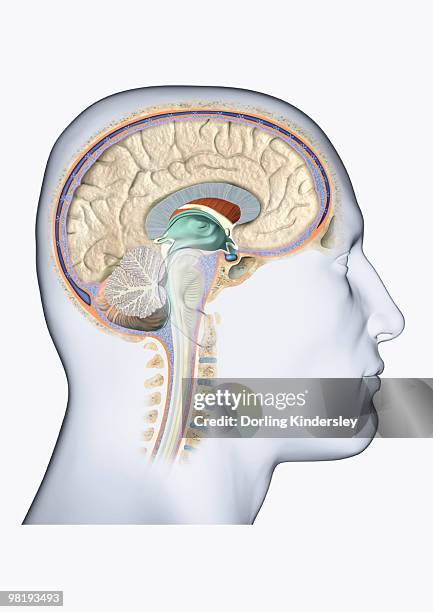
digital illustration of head in profile showing cross section of brain, neck vertebra and spine - 丘腦 幅插畫檔、美工圖案、卡通及圖標腦下垂體腺
digital illustration of human brain with sound entering via brain stem and thalamus to auditory cortex - 丘腦 幅插畫檔、美工圖案、卡通及圖標
digital illustration of head in profile showing brain with basal ganglia, thalamus and frontal cortex highlighted - 丘腦 幅插畫檔、美工圖案、卡通及圖標
cross section digital illustration of brain highlighting cerebral cortex, thalamus, and brain stem - 丘腦 幅插畫檔、美工圖案、卡通及圖標
digital illustration of dorsolateral prefrontal cortex highlighted in green, anterior cingulate cortex and thalamus in human brain - 丘腦 幅插畫檔、美工圖案、卡通及圖標前額葉皮質
digital illustration of signal travelling from spinal cord through brain stem, connecting nerve fibre with third-order neuron in thalamus to somatosensory cortex - 丘腦 幅插畫檔、美工圖案、卡通及圖標
digital illustration of head in profile showing areas of brain connected to emotion - 丘腦 幅插畫檔、美工圖案、卡通及圖標
digital illustration of areas of activity during rem sleep in human brain highlighted in red and green - 丘腦 幅插畫檔、美工圖案、卡通及圖標前額葉皮質, 杏仁核