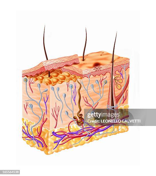
medical illustration cross section of human skin layers - 19th century - 乳突 幅插畫檔、美工圖案、卡通及圖標皮膚科, 插圖, 生物醫學圖解
Royal spoonbill , feeding by sweeping bill from side to side in shallow muddy water and snapping up aquatic organisms that it feels. Narran Lake, New...
The Macula Whitish Oval Spot Is Located On The Retina, On The Lower, Outer Side Of The Optical Disk. Photograph Of The Back Wall Of Eye.
The Macula Whitish Oval Spot Is Located On The Retina, On The Lower, Outer Side Of The Optical Disk. Photograph Of The Back Wall Of Eye.
Glaucoma Of Optical Disk. Ocular Hypertonia Is Caused By Poor Circulation In Intra Ocular Aqueous Humor. Modification Of The Optical Disk And Field...青光眼
Glaucoma Of Optical Disk. Ocular Hypertonia Is Caused By Poor Circulation In Intra Ocular Aqueous Humor. Modification Of The Optical Disk And Field...青光眼
Glaucoma Of Optical Disk. Ocular Hypertonia Is Caused By Poor Circulation In Intra Ocular Aqueous Humor. Modification Of The Optical Disk And Field...青光眼
Worldwide Distribution Except For Canada. Nephron. Representation Of The Structure Of The Nephron With Its Glomerulus Brown Sphere At The Top In The...
Glaucoma. Illustration Of A Glaucomatous Eye With Two Highlighted Areas Showing Open Angle Glaucoma And A Glaucomatous Blind Spot.
Glaucoma Iridial Angle, Open And Closed Angles. Anterior Part Of The Eye: Comparison Between What Occurs First In A Healthy Eye, Then In An Eye...
Angle Closure Glaucomaillustration Of Angle Closure Glaucoma: The Trabecular Meshwork Is Blocked By The Pupil, Preventing The Aqueous Humor From...
Cross Section Of The Skin And Its Various Constituent Elements. Illustration Of The Skin, Including Its Various Layers: Epidermis, Dermis,...
Illustration Of The Tongue Which Maps The Various Flavor Areas: Bitterness Green Sourness Pink Sweetness Yellow Saltiness Blue
Anatomic Model Of The Human Right Eye Lateral View Of A Median Section. The Eyeball Is Composed Of An Outer Fibrous Coat, Consisting Of The Sclera A...
Model Of The Intern Anatomy Of The Kidney Of An Adult Human Body Anterior View Of A Frontal Section. The Kidney, Wrapped In A Fibrous Capsule In...
Model Of The Anatomy Of The Urinary Tract Of A Female Adult Human Body, Anterior View. The Deep Organs Can Be Seen Here Thanks To The Removal Of The...
Model Of The Internal Anatomy Of The Urinary Tract Of An Adult Human Body Of Indeterminate Sex, Anterior View. The Deep Organs Can Be Seen Here...
Model Of The Internal Anatomy Of The Right Kidney Of An Adult Human Body Anterior View Of A Frontal Section. The Kidney Filters The Blood That It...塑膠
Model Of The Internal Anatomy Of The Right Kidney Of An Adult Human Body Anterior View Of A Frontal Section. The Kidney Filters The Blood, That It...塑膠
Anatomic Model Of The Skin Frontal Section. The Integumentary System Includes The Skin And Its Appendages Hairs, Glands, Muscles And Nerves. The Skin...蜂窩性組織炎
Anatomic Model Of The Skin Frontal Section. The Integumentary System Includes The Skin And Its Appendages Hairs, Glands, Muscles And Nerves. The Skin...
Anatomic Model Of The Skin Frontal Section. The Integumentary System Includes The Skin And Its Appendages Hairs, Glands, Muscles And Nerves, Depicted...毛囊, 蜂窩性組織炎
Model Of The Internal Anatomy Of The Trunk Of An Adult Human Body Of Indeterminate Sex, Anterior View Of The Kidney Region. The Deep Organs Can Be...
Photo Essay From The University Hospital Of Bordeaux. Cardiologic Hospital Of Haut Leveque. Department Of Diabetology. Examination Of Retinography...糖尿病
Model Of The Internal Anatomy Of The Trunk Of An Adult Human Body Of Indeterminate Sex, Anterior View. The Deep Organs Can Be Seen Here Thanks To The...
Illustration Of A Transversal Section Of The Eye Eyelash, Aqueous Humor, Cornea, Colored Iris, Vitreous Humor, Fovea, Sclerotic And Right Upper And...
Illustration For Children Representing The Area Of Taste In Yellow The Sweetness, In Blue The Saltiness, In Red The Acidity And In Green The...
The Eye. Anatomy Of The Right Eye Horizontal Cutaway View Showing The Main Structures Of The Eye, Including The Retina And Its Vascularization, The...
The Eye. Anatomy Of The Right Eye Horizontal Cutaway View Showing The Main Structures Of The Eye, Including The Retina And Its Vascularization, The...
Anatomy Of The Eye. Representation Of The Anatomy Of The Eye In Saggital View. La Macula Or Yellow Spot, Here In Dark Blue Is Located At The Center...
Anatomy Of The Eye. Representation Of The Anatomy Of The Eye In Saggital View. La Macula Or Yellow Spot, Here In Dark Blue Is Located At The Center...