Photomicrograph of lung tissue with various histopathologic changes indicative of Toxic Shock Syndrome , taken during an autopsy, 1984. Image...差異
Magnified 1000X, this micrograph depicts Bacteroides fragilis subsp. Fragilis bacteria that had been grown in Schaedler's broth, and processed using...
Acne and its treatments, the sebaceous gland is in hypersecretion. Cut of skin of the epidermis and dermis. Normal hair follicle and acne pimple in...
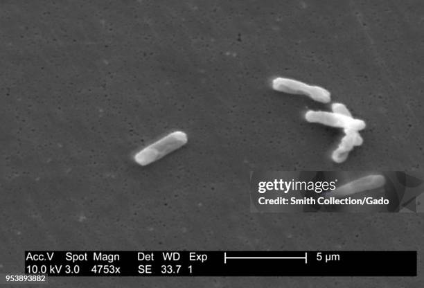
Gram-positive Clostridium difficile bacteria revealed in the stool sample micrograph film, 2004. Image courtesy Centers for Disease Control / Lois S....細菌
Gram-positive Clostridium difficile bacteria revealed in the stool sample micrograph film, 2004. Image courtesy Centers for Disease Control / Lois S....
Gram-positive Clostridium difficile bacteria revealed in the stool sample micrograph film, 2004. Image courtesy Centers for Disease Control / Lois S....
Gram-positive Clostridium difficile bacteria revealed in the stool sample micrograph film, 2004. Image courtesy Centers for Disease Control / Lois S....
Gram-positive Clostridium difficile bacteria revealed in the stool sample micrograph film, 2004. Image courtesy Centers for Disease Control / Lois S....
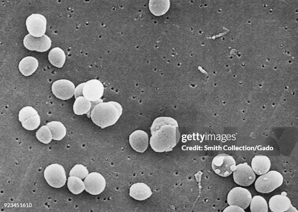
Gram-positive Staphylococcus aureus bacteria revealed in a scanning electron microscopic image, 2003. Image courtesy Centers for Disease Control .
Photomicrograph of Staphylococcus aureus bacteria, a causative agent of Toxic Shock Syndrome , on a Gram-stained smear, 1984. Image courtesy CDC.
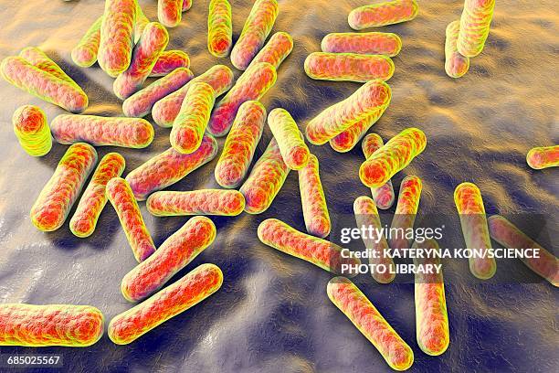
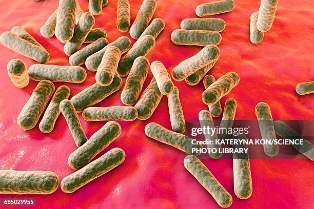
mycobacterium chelonae bacteria, colored scanning electron micrograph (sem) - 革蘭氏陽性菌 個照片及圖片檔細菌, 掃描電子顯微拍攝, 食物中毒
This micrograph depicts the gram-positive bacterium Propionibacterium acnes grown in thioglycollate medium at 48 hours, 1972. Propionibacterium acnes...
This micrograph depicts the gram-positive bacterium Bifidobacterium eriksonii, 1972. B. Eriksonii, also called Actinomyces eriksonii, is an anaerobic...雙歧桿菌, 細菌
This micrograph depicts histopathologic changes due to the gram-positive organism, Actinomyces israelii, 1971. Using a modified Fite-Faraco stain, a...
Pictured is a Lowenstein-Jensen Slant culture of Nocardia asteroids at 37°C, 1969. The bacterial complex Nocardia asteroids is a serious threat to...
This photomicrograph depicts the histopathologic changes associated with nocardiosis of the lung, 1973. 80% of the cases of nocardiosis show clinical...
This micrograph depicts Streptococcus pneumoniae bacteria in cerebral spinal fluid using FA staining technique, 1964. The bacterium Streptococcus...
This is a plate culture of the bacteria Nocardia asteroides grown on 7H10 agar medium at 37° C, 1969. The bacterial complex Nocardia asteroids is a...
This is a plate culture of the bacteria Nocardia asteroides grown on 7H10 agar medium at 37° C, 1969. The bacterial complex Nocardia asteroids is a...
This is a plate culture of the bacteria Nocardia asteroides grown on 7H10 agar medium at 37° C, 1969. The bacterial complex Nocardia asteroids is a...細菌, 培養皿
This is a plate culture of the bacteria Nocardia asteroides grown on 7H10 agar medium at 37° C, 1969. The bacterial complex Nocardia asteroids is a...
This is a plate culture of the bacteria Nocardia asteroides grown on 7H10 agar plates at 37° C, 1969. The bacterial complex Nocardia asteroids is a...
Magnified 560X, this photomicrograph showed the presence of Pneumocystis jirovecii, formerly known as Pneumocystis carinii fungal organisms using an...抗體
These slant cultures growing Actinomyces viscosus, reveal the oxygen requirements of this bacterium, 1971. Each culture represents the following...
This methylamine silver stained brain abscess tissue sample shows histopathologic changes due to A. Naeslundii bacteria, 1971. Pathology associated...
These slant cultures growing Actinomyces viscosus, reveal the oxygen requirements of this bacterium, 1971. Each culture represents the following...發現
This micrograph depicts the gram-positive bacterium Propionibacterium acnes grown in blood agar for 48 hours, 1972. Propionibacterium acnes is a very...細菌
Agar plate culture of Actinomyces sp. Strain X-573, at 7 days, incubated anaerobically, 1963. Actinomyces sp. Normally found in the oral cavity is an...
Agar plate culture of Actinomyces sp. Strain X-573, at 7 days, incubated anaerobically, 1963. Actinomyces sp. Normally found in the oral cavity is an...
Agar plate culture of Actinomyces sp. Strain X-407, at 7 days, incubated anaerobically, 1963. Actinomyces sp. Normally found in the oral cavity is an...
Agar plate culture of Actinomyces sp. Strain X-407, at 7 days, incubated anaerobically, 1963. Actinomyces sp. Normally found in the oral cavity is an...
Agar plate culture of Actinomyces sp. Strain X-573, incubated anaerobically, 1963. Actinomyces sp. Normally found in the oral cavity is an...
Plate culture of Actinomyces sp. Strain- X616, at 72hrs of growth, 1963. Actinomyces sp. Normally found in the oral cavity, is an opportunistic...
Plate culture of Actinomyces sp. A gram-positive opportunistic pathogen that is normally found in the oral cavity, 1963. Actinomyces spp. Are usually...
Plate culture of Actinomyces sp. A Gram positive opportunistic pathogen that is normally found in the oral cavity, 1963. Actinomyces spp. Are usually...
This micrograph depicts Nocardia brasiliensis found in a purulent exudate from a skin lesion using gram-stain, 1973. Bacterial members of the genus...
This is a photomicrograph of Dermatophilus congolensis bacteria using a Giemsa stain, 1972. Dermatophilus congolensis is a gram-positive,...細菌
This micrograph demonstrates a clustering of Dermatophilus congolensis bacteria using a Giemsa stain, 1973. D. Congolensis is a motile, nonacid-fast,...
This tissue specimen shows signs of Pitted Keratolysis due to a Dermatophilus sp. Infection, using a PAS stain, 1972. Pitted keratolysis is a skin...