back view of human skeleton with nervous system, arteries and veins. - thoracic vertebrae 幅插畫檔、美工圖案、卡通及圖標
back view of human skeleton with nervous system, arteries and veins. - thoracic vertebrae 幅插畫檔、美工圖案、卡通及圖標
medical illustration of human body showing skeletal system, arteries, veins, and nervous system. - thoracic vertebrae 幅插畫檔、美工圖案、卡通及圖標
medical illustration showing inflammation and pain in human back area, perspective view. - thoracic vertebrae 幅插畫檔、美工圖案、卡通及圖標
medical illustration showing inflammation and pain in human neck area, perspective view. - thoracic vertebrae 幅插畫檔、美工圖案、卡通及圖標
medical illustration of human brain and brain stem, perspective view. - thoracic vertebrae 幅插畫檔、美工圖案、卡通及圖標
three dimensional view of female upper back and skeletal system. - thoracic vertebrae 幅插畫檔、美工圖案、卡通及圖標人類骨架
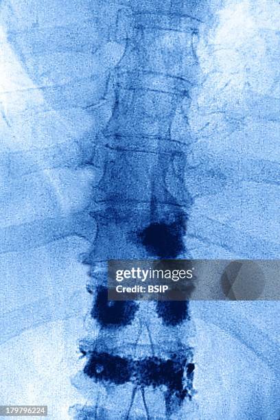
Pagets Disease, X-Ray, Osteoplastic Intervention To Repair Lumbar Vertebrae By Injecting Prosthetic Cement In The Vertebral Disks To Prevent Medullar...X光片, 椎間盤突出
Metastasis, Thoracic Vertebrae, Sagital MRI Image Of The Thoracic Spine Shows A Malignant Tumor Of A Intervertebral Disc With Medullar Compression.人體骨骼, 癌症, 癌擴散
Osseous Metastasis On A Dorsal Vertebra, Secondary Malignant Tumor. Vertebral X Ray, Thorax In Front View.癌症, X光片, 癌擴散
Osseous Metastasis On A Dorsal Vertebra, Secondary Malignant Tumor. Vertebral X Ray, Thorax In Front View.
Scanner Of The Lumbar Vertebrae Anterior View. Thresholding To Obtain The Visualization Of The Bones. Last Thoracic Vertebra T12, Lumbar Vertebrae L1...
Osteoplastic Intervention To Repair Lumbar Vertebrae By Injecting Prosthetic Cement In The Vertebral Disks To Prevent Medullar Compression By Spinal...
Osteoplastic Intervention To Repair Lumbar Vertebrae By Injecting Prosthetic Cement In The Vertebral Disks To Prevent Medullar Compression By Spinal...
Osteoplastic Intervention To Repair Lumbar Vertebrae By Injecting Prosthetic Cement In The Vertebral Disks To Prevent Medullar Compression By Spinal...
Osteoplastic Intervention To Repair Lumbar Vertebrae By Injecting Prosthetic Cement In The Vertebral Disks To Prevent Medullar Compression By Spinal...
This Sagital Mri Image Of The Thoracic Spine Shows A Malignant Tumor Of A Intervertebral Disc With Medullar Compression.胸部
The superior mediastinum is that portion of the interpleural space which lies between the manubrium sterni in front, and the upper thoracic vertebrae...
Posterior Medistinum, view of trachea, bronchi, lungs, and heart from behind, 1903. From 'Surgical Anatomy: The Treatise of the Human Anatomy and Its...
The quadrus lumborum muscle is a thin, quadrilateral muscle, 1903. From 'Surgical Anatomy: The Treatise of the Human Anatomy and Its Applications to...
The superior mediastinum is that portion of the interpleural space which lies between the manubrium sterni in front, and the upper thoracic vertebrae...
The cervical sympathetic nerves are a paired bundle of nerve fibers that run from the base of the skull to the coccyx, 1903. From 'Surgical Anatomy:...
Illustration from 'Surgical Anatomy: The Treatise of the Human Anatomy and Its Applications to the Practice of Medicine and Surgery, volume III'...
Illustration from 'Surgical Anatomy: The Treatise of the Human Anatomy and Its Applications to the Practice of Medicine and Surgery, volume III'...
Illustration from 'Surgical Anatomy: The Treatise of the Human Anatomy and Its Applications to the Practice of Medicine and Surgery, volume III'...
Illustration from 'Surgical Anatomy: The Treatise of the Human Anatomy and Its Applications to the Practice of Medicine and Surgery, volume III'...
Illustration from 'Surgical Anatomy: The Treatise of the Human Anatomy and Its Applications to the Practice of Medicine and Surgery, volume III'...
Fixation by screws and plates on the sixth, seventh cervical vertebrae and the first thoracic vertebra, after vertebral dislocation, visualized by...
Fixation by screws and plates on the sixth, seventh cervical vertebrae and the first thoracic vertebra, after vertebral dislocation, visualized by...
Fixation by screws and plates on the sixth, seventh cervical vertebrae and the first thoracic vertebra, after vertebral dislocation, visualized by...
Fixation by screws and plates on the sixth, seventh cervical vertebrae and the first thoracic vertebra, after vertebral dislocation, visualized by...
Rotational dislocation at the junction of the thoracic and lumbar vertebrae, visualized by an AP chest radiograph.
Rotational dislocation at the junction of the thoracic and lumbar vertebrae, visualized by an AP chest radiograph.
Rotational dislocation at the junction of the thoracic and lumbar vertebrae, visualized by an AP chest radiograph.
Rotational dislocation at the junction of the thoracic and lumbar vertebrae, visualized by an AP chest radiograph.