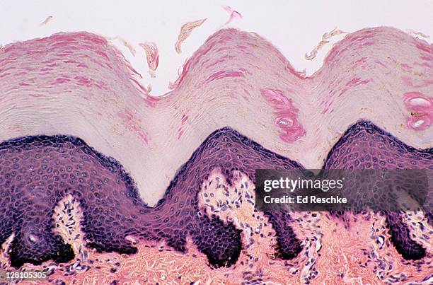
skin. desquamation (sloughing of cells) from the epidermis, thick skin, human, 100x at 35mm. shows: epidermal layers (stratum: corneum, granulosum, spinosum, basale), dermis, sweat gland ducts and desquamating cells sloughing off the surface. - 光學顯微圖 個照片及圖片檔細胞, 有層次
Micrograph showing the histologic changes in human skin infected with smallpox, prepared with hematoxylin-eosin stain, 1975. .
hyaline cartilage; tracheal cartilage, supporting connective tissue, 250x at 35mm. shows: chondrocytes (cartilage cells), and matrix (intercellular material, non-living). - 光學顯微圖 個照片及圖片檔科學顯微圖片, 細胞, 人類氣管
epididymis. pseudostratified columnar epithelium with stereocilia, 250x at 35mm. shows: epithelium with stereocilia and the lumen with spermatozoa. sperm are temporaily stored in the epididymis. - 光學顯微圖 個照片及圖片檔
Photomicrograph of the malaria parasite Plasmodium Ovale growing as a double trophozoite in one red blood cell and a single trophozoite, on a thin...血球
Aluminium 4% copper . Aluminium 4% copper . Grains of solid solution of copper in aluminium. Light micrograph in bright field. Magnification 500x.
colored light micrograph of a section through hairs of the human skin - 光學顯微圖 個照片及圖片檔科學顯微圖片, 毛囊, 彩色影像
Micrograph of a macroconidium and some microconidia of the fungus Trichophyton rubrum var, 1973. Rodhaini. Image courtesy CDC/Dr. Libero Ajello. .
This micrograph depicts the gram-positive bacterium Bifidobacterium eriksonii, 1972. B. Eriksonii, also called Actinomyces eriksonii, is an anaerobic...雙歧桿菌, 細菌
This micrograph depicts the histopathologic changes associated with histoplasmosis of the spleen using Gridley stain, 1964. Positive histoplasmin...
Magnified 1125X, this thin-film, Giemsa-stained photomicrograph revealed the presence of a growing Plasmodium ovale trophozoite, with a ring nucleus....分析