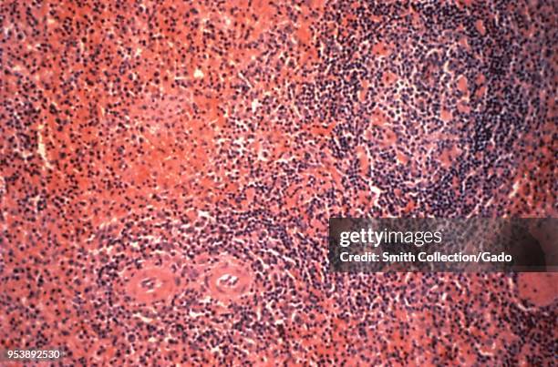
Liver tissue revealed in the micrograph image from a Hantavirus pulmonary syndrome patient, 1994. Image courtesy Centers for Disease Control .漢坦病毒
Splenic tissue revealed in the micrograph image from a Hantavirus pulmonary syndrome patient, 1994. Image courtesy Centers for Disease Control .漢坦病毒
Lymph node cortex revealed in the micrograph image from a Hantavirus pulmonary syndrome patient, 1994. Image courtesy Centers for Disease Control .漢坦病毒
Atypical enlarged lymphocyte revealed in the blood smear micrograph from an HPS patient, 1994. Image courtesy Centers for Disease Control .漢坦病毒
Presence of herpes simplex virus in a Tzanck test specimen revealed in the micrograph film, 1975. Image courtesy Centers for Disease Control / Joe...單純皰疹病毒, 皰疹
Histopathologic changes found in the case of Kaposi's sarcoma lesions revealed in the skin biopsy micrograph film, 1981. Image courtesy Centers for...
Eukocytosis micrograph revealing an elevated number of white blood cells, 1974. Image courtesy CDC/Dr. Candler Ballard.
White blood cells , a stack of platelets, and red blood cells revealed in the photomicrograph of a thick film Giemsa-stained blood smear sample,...紫色
Babesia microti and Plasmodium berghei revealed in red blood cells using Giemsa stained, 1972. Image courtesy Centers for Disease Control / Dr George...