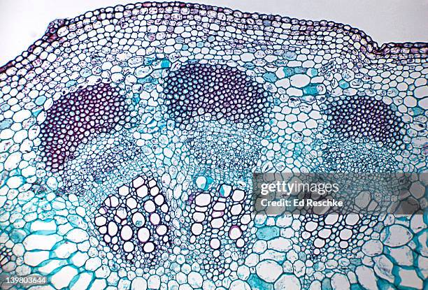
sunflower. helianthus stem. vascular bundles. supporting tissue sclerenchyma. dicot. 100x at 35mm - 厚壁組織 個照片及圖片檔
x-section of indian corn stem (zea mays). shows vascular bundles (xylem and phloem), sclerenchyma, sieve tubes, and companion cells. (100x) - 厚壁組織 個照片及圖片檔科學顯微圖片
triticum (wheat). stem cross section. monocot. shows: vascular bundle, xylem, phloem, shlorenchyma, epidermis. 50x at 35mm - 厚壁組織 個照片及圖片檔基本組織, 細胞
monocot stem (cross section) with scattered vascular bundles--greenbrier (smilax sp) 8x - 厚壁組織 個照片及圖片檔
grass leaf anatomy, lasiagrostis calamagrostis and festuca porcii leaf cross sections - 厚壁組織 幅插畫檔、美工圖案、卡通及圖標
Paléontologie française, Paris, Chez l'auteur, 1840-1894, France, paleontology, Jurassic, Tertiary, Cretaceous, paleobotany, mollusk fossil, sea...
The image presents oak xylem tissue in the transversal cross-section of the stalk, photographed through the microscope in polarized light at a...
The image presents nettle tissues in the transversal section of the stalk, photographed through the microscope in polarized light at a magnification...
The image presents nettle tissues in the transversal cross-section of the stalk, photographed through the microscope in polarized light at a...
The image presents nettle tisues in the transversal cross-section of the stalk, photographed through the microscope in polarized light at a...
The image presents nettle tissues in the stalk in transversall cross-section, photographed through the microscope in polarized light and dark field...
zea mays (indian corn). stem cross section. xylem, phloem, sclerenchyma. epidermis, parenchyma. 50x h - 厚壁組織 個照片及圖片檔
stem cross section. wheat (triticum), herbaceous monocot, 100x. shows: a vascular bundle, xylem, phloem, sclerenchyma, chlorenchyma, epidermis, parenchyma, and bundle sheath extension. - 厚壁組織 個照片及圖片檔薄壁組織
stem cross section. wheat (triticum), herbaceous monocot, 50x. shows: 2 vascular bundles, xylem, phloem, epidermis, sclerenchyma, parenchyma, chlorenchyma (green areas near surface), and hollow pith. - 厚壁組織 個照片及圖片檔木髓
stem cross section. sunflower (helianthus), herbaceous dicot, 50x. shows a single vascular bundle. vascular bundles are arranged in a ring. shows: xylem, phloem, cambium, sclerenchyma (bundle cap), and surrounding parenchyma. - 厚壁組織 個照片及圖片檔幹細胞, 背景 - 主題
stem cross section. sunflower (helianthus), herbaceous dicot, 25x. vascular bundles arranged in a ring. shows: xylem, phloem, cambium, sclerenchyma (bundle cap), epidermis, cortex, and pith. - 厚壁組織 個照片及圖片檔木髓
stem cross section. corn (zea), herbaceous monocot, 100x. shows a single vascular bundle with: xylem, phloem, companion cells, sieve tubes, and sclerenchyma. - 厚壁組織 個照片及圖片檔
stem cross section. corn (zea), herbaceous monocot, 25x. shows: scattered vascular bundles typical of monocots, xylem, phloem, sclerenchyma, pith, and epidermis. - 厚壁組織 個照片及圖片檔顯微鏡, 科學顯微圖片
helianthus, sunflower stem, single vascular bundle, shows xylem, phloem, cambium, sclerenchyma 50x at 35mm - 厚壁組織 個照片及圖片檔室內
microscopic view of cells of broad bean plant. immunofluorescent photomicrograph, organs samples, histological examination, histopathology on the microscope - 厚壁組織 個照片及圖片檔微距攝影, 顯微鏡, 抽象
dicotyledon leaf cross-section, vascular bundle, vascular tissue, immunofluorescent photomicrograph, organs samples, histological examination, histopathology on the microscope - 厚壁組織 個照片及圖片檔
pteridium aquilinum dennstaedtiaceae. immunofluorescent photomicrograph, organs samples, histological examination, histopathology on the microscope - 厚壁組織 個照片及圖片檔基本組織, 分生組織的, 顯微鏡
triticum (wheat) stem, monocot. shows vascular bundle, xylem, phloem, sclerenchyma, chlorenchyma & epidermis. 100x at 35mm - 厚壁組織 個照片及圖片檔細胞
vascular bundles and plant anatomy in a herbaceous dicot stem---sunflower (helianthus) cross section, 50x - 厚壁組織 個照片及圖片檔
vascular bundle of a herbaceous monocot stem (corn, zea mays) xylem, phloem, companion cells, bundle sheath, 100x - 厚壁組織 個照片及圖片檔
herbaceous monocot stem (corn, zea mays) vascular bundle--xylem, phloem, companion cells, bundle sheath, 100x - 厚壁組織 個照片及圖片檔