cross section biomedical illustration of human digestive system connected to esophagus - 唾腺 幅插畫檔、美工圖案、卡通及圖標
stratified columnar epithelium. duct of submandibular salivary gland, 250x shows: tall columnar cells (surface) and other cell sizes. two or more layers--surface cells columnar. - 唾腺 個照片及圖片檔上皮組織
parotid salivary gland. 250x. shows: serous cells (dark, granular cells) and a duct. the serous cells show secretion granules which are a source of enzymes (salivary amylase). human. - 唾腺 個照片及圖片檔口水, 細胞
muscle cross section in microscopy , education anatomy and histopathology of in laboratory. - 唾腺 個照片及圖片檔
education anatomy and histopathology of tongue under the microscopic in laboratory. - 唾腺 個照片及圖片檔抽象, 背景 - 主題
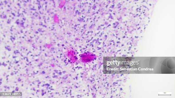
microscope of adenoid cystic carcinoma, rare type of cancer exist in many different body sites. this tumor occurs in the salivary glands, - 唾腺 個照片及圖片檔癌症, 血球
The digestive anatomy of the newborn in this illustration shows the organs from the oral cavity to the rectum. The great peculiarity of the anatomy...兒童
This confocal microscope image shows a cross section of a tick salivary gland infected with Langat virus . Two rounded structures on the right,...蜱 - 動物, 口水
normal human submandibular salivary gland, 100x at 35mm. showing mucous cells (clear), serous cells (dark), and a duct. mixed gland. serous cells show secretion granules; source of enzymes. - 唾腺 個照片及圖片檔
normal human submandibular salivary gland, 100x at 35mm. showing mucous cells (clear), serous cells (dark, with secretions granules), and a duct. mixed gland. the dark secretion granules in the serous cells are the source of enzymes. - 唾腺 個照片及圖片檔
old engraved illustration of anatomy of the paranasal sinuses (spaces between the bones around the nose) - 唾腺 個照片及圖片檔心型, 古董, 人體
serous glands (with zymogen granules) in the parotid salivary gland---serous cells and duct, 250x - 唾腺 個照片及圖片檔