cross section - xylem is a type of tissue in vascular plants that transports water and some nutrients. scientific research. plant tissue structureimmunofluorescent photomicrograph, organs samples, histological examination, histopathology on the microscope - 形成層 個照片及圖片檔細胞, 分析, 顯微鏡
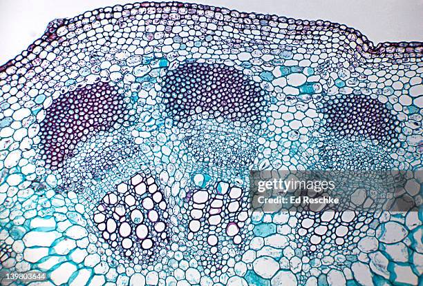
anatomy of a herbaceous dicot stem--sunflower (helianthus annus) with 2 vascular bundles, 50x - 形成層 個照片及圖片檔人體構造
Portugal, cork industry, men bringing cork tissue aboard, date unknown, probably 1936, photo by Eric Borchert, picture belongs to a series of...
Portugal, cork industry, typical boat loaded with cork tissue, date unknown, probably 1936, photo by Eric Borchert, picture belongs to a series of...
Portugal, cork industry, men pushing a cart loaded with hot and dampish cork, date unknown, probably 1936, photo by Eric Borchert, picture belongs to...
Dan West, an entomologist with the Colorado State Forest Service, uses a draw knife to peel bark off of local ash trees looking for the Emerald Ash...
Cassey Anderson, left, a project manager at CSU Extension and Micaela Truslove, right, the EAB program coordinator for the Colorado Department of...
cross section of laurel clockvine, thunbergia laurifolia, rhynchosia phaseoloides, trumpet vine, tecoma radicans - 形成層 幅插畫檔、美工圖案、卡通及圖標
deciduous tree branch, angiosperm stem, schematic cross section showing wood and bark tissues - 形成層 幅插畫檔、美工圖案、卡通及圖標
this botanical engraving illustrates the internal cellular structure of a one-year-old twig of the blackcurrant (ribes nigrum) - 形成層 幅插畫檔、美工圖案、卡通及圖標
abstract background from hura crepitans, the tropical sandbox tree grown in central vietnam - 形成層 個照片及圖片檔
abstract background from hura crepitans, the tropical sandbox tree grown in central vietnam - 形成層 個照片及圖片檔
abstract background from hura crepitans, the tropical sandbox tree grown in central vietnam - 形成層 個照片及圖片檔
maple tree (acer) trunk cross sections showing medullary ray cells - 19th century - 形成層 幅插畫檔、美工圖案、卡通及圖標植物細胞, 細胞
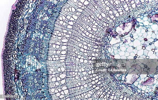
3 yr stem tilia (basswood) shows pith annual rings (3), cambium phloem, rays, periderm, cork. 10x h - 形成層 個照片及圖片檔
tilia (3 year old stem). shows: cortex, xylem, phloem, pith, rays, cambium. 50x basswood.woody dicot stem h - 形成層 個照片及圖片檔
tilia. 3 year stem. woody dicot xylem, phloem, pith, fibers, phloem ray, annual rings (3), cambrium vascular cambium, springwood, summer wood. 5x - 形成層 個照片及圖片檔木髓
woody dicot stem. tilia (basswood), 3 year stem, 5x. shows: 3 annual rings, xylem, phloem, rays, pith, cambium, and periderm. - 形成層 個照片及圖片檔
woody dicot stem. tilia (basswood), 2 year stem, 2.5x. shows: 2 annual rings, xylem, phloem, rays, pith, cambium, periderm - 形成層 個照片及圖片檔
woody dicot stem. tilia (basswood), 2 year stem, 25x. shows: xylem, phloem, pith, bark, cork, vascular cambium and two annual rings. - 形成層 個照片及圖片檔科學顯微圖片, 幹細胞, 木髓
woody dicot stem. tilia (basswood), 2 year stem, 10x. shows: 2 annual rings, xylem, phloem, rays, pith, cambium, periderm - 形成層 個照片及圖片檔幹細胞
woody dicot stem. tilia (basswood), 2 year stem, 7x. shows: xylem, phloem, two annual rings, pith, rays, vascular cambium, periderm - 形成層 個照片及圖片檔木質部, 顯微鏡, 科學顯微圖片
woody dicot stem. tilia (basswood), 1st year stem, 10x. shows: xylem, phloem, pith, cortex, bark, cork, rays, vascular cambium, and one annual ring. - 形成層 個照片及圖片檔顯微鏡
stem cross section. sunflower (helianthus), herbaceous dicot, 50x. shows a single vascular bundle. vascular bundles are arranged in a ring. shows: xylem, phloem, cambium, sclerenchyma (bundle cap), and surrounding parenchyma. - 形成層 個照片及圖片檔幹細胞, 背景 - 主題
stem cross section. sunflower (helianthus), herbaceous dicot, 25x. vascular bundles arranged in a ring. shows: xylem, phloem, cambium, sclerenchyma (bundle cap), epidermis, cortex, and pith. - 形成層 個照片及圖片檔木髓
stem cross section. basswood (tilia), 3 year stem, woody dicot, 25x. shows: 3 annual rings (with springwood & summerwood), xylem, phloem, pith, cambium, phloem ray, cortex, periderm. - 形成層 個照片及圖片檔木質部
stem cross section. basswood (tilia), 1st year stem, woody dicot, 25x. shows: xylem, phloem, cambium, rays, pith, cork, and collenchyma. - 形成層 個照片及圖片檔顯微鏡
helianthus, sunflower stem, single vascular bundle, shows xylem, phloem, cambium, sclerenchyma 50x at 35mm - 形成層 個照片及圖片檔室內
The exposed cambium layer beneath the burned bark of a giant sequoias that was damaged by the Pier Fire in 2017 in the Sequoia National Forest near...
microscopic view of cells of broad bean plant. immunofluorescent photomicrograph, organs samples, histological examination, histopathology on the microscope - 形成層 個照片及圖片檔微距攝影, 顯微鏡, 抽象
dicotyledon leaf cross-section, vascular bundle, vascular tissue, immunofluorescent photomicrograph, organs samples, histological examination, histopathology on the microscope - 形成層 個照片及圖片檔
pteridium aquilinum dennstaedtiaceae. immunofluorescent photomicrograph, organs samples, histological examination, histopathology on the microscope - 形成層 個照片及圖片檔基本組織, 分生組織的, 顯微鏡