aerial view/solar cells float clean energy that will be the energy of the future. - 植物細胞 個照片及圖片檔太陽能電池板, 農場
giant splenic artery aneurysm seen on mip reconstruction on ct image (cat scan) - 植物細胞 個照片及圖片檔電腦斷層掃描, 動脈瘤, 脾臟
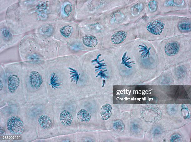
plant mitosis, anaphase, onion (allium) root tip. (magnification x400). the chromosomes have moved to opposite ends (poles) of the cell. quadruple stain. - 植物細胞 個照片及圖片檔顯微鏡, 細胞, 洋蔥
dicot stem. helianthus (sunflower) vascular bundles (ring), phloem, xylem, pith cortex, epidermis. 10x at 35mm. vascular bundles arranged in a ring. organization of primary tissues. - 植物細胞 個照片及圖片檔圓形, 維管束
microscopic view of cells of broad bean plant. immunofluorescent photomicrograph, organs samples, histological examination, histopathology on the microscope - 植物細胞 個照片及圖片檔微距攝影, 顯微鏡, 抽象
illustration of the structure of a plant cell, including nucleus, nucleolus, ribosome, endoplasmatic - 植物細胞 幅插畫檔、美工圖案、卡通及圖標
manipulated image of palm leaf in symmetrical vibrant green and yellow colors and clean lines - 植物細胞 個照片及圖片檔萬花筒式樣, 動作, 樹葉
single tropical leaf showing graceful red outline of leaf edge on black background - low key - 植物細胞 個照片及圖片檔黑色的背景, 大自然, 光合作用
Illustration of a plant cell's primary cell wall consisting of cellulose, hemicellulose, proteins, pectins and calcium.
stem cross section of brazilian waterweed (egeria densa) showing cortex, pith and vascular tissue. magnified 60x (ref. at 35 mm film frame) - 植物細胞 個照片及圖片檔顯微鏡, 細胞, 剪裁圖
old engraved illustration of powdery mildews and moulds of leaves, paper, straw, and herbarium specimens. the plate compares fungal fruiting bodies, sporangia, conidia, mycelium, and infected materials. - 植物細胞 個照片及圖片檔
old engraved illustration of rust and smut stages on burnet, barren strawberry, rose, bramble, raspberry, meadowsweet, and other host plants. the plate traces infected leaves, telial stages, spores, and germination structures. - 植物細胞 個照片及圖片檔
old engraved illustration of powdery mildew structures of sphaerotheca, phyllactinia, uncinula, microsphaeria, oidium, and erysiphe. the plate compares fruiting bodies, sporangia, appendages, conidia, and infected leaves. - 植物細胞 個照片及圖片檔
old engraved illustration of white rust and downy mildew structures of cystopus and peronospora. the plate diagrams conidia, mycelium, oogonia, zoospores, and germination stages. - 植物細胞 個照片及圖片檔
old engraved illustration of aecidia and ripe spermogonia of aecidium grossulariae and aecidium euphorbiae. the two cross sections identify cluster-cups, spermogonia, spermatia, and sterigmata. - 植物細胞 個照片及圖片檔
abstract green liquid bubbles with transparent glossy texture in macro view. futuristic 3d background inspired by biotechnology, skincare serum, microscopic cells, eco science and natural cosmetics. organic fluid shapes for beauty and biological research. - 植物細胞 個照片及圖片檔細胞, 環境保護, 抽象
Botanical and Physiological Memoirs London Printed for the Ray Society 1853 Plant Physiology Fragilariaceae Proto coccus pluvialis Rejuvenescence...
this botanical engraving illustrates the internal cellular structure of a one-year-old twig of the blackcurrant (ribes nigrum) - 植物細胞 幅插畫檔、美工圖案、卡通及圖標
old engraved illustration of cartilage cells, primarily chondrocytes - the specialized cells responsible for producing and maintaining the cartilaginous matrix, which consists of collagen, proteoglycans and water - 植物細胞 個照片及圖片檔
old engraved illustration of yellow fibro cartilage, elastic cartilage or yellow elastic cartilage, which contains a dense network of yellow elastic fibers alongside collagen fibers - 植物細胞 個照片及圖片檔