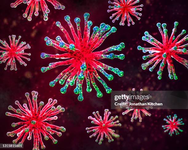
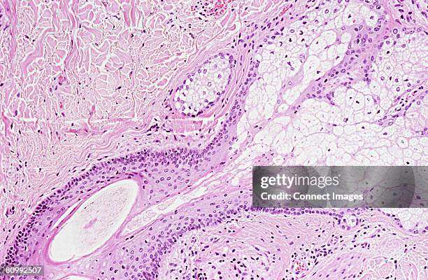
bacteria, cells, molecules view in a microscope. close-up. 3d render. healthcare and medicine. - 科學顯微圖片 個照片及圖片檔培養皿, 顯微鏡, 細菌
cardiac muscle shows syncytium (interconnected cell network), striations, intercalated disks & nuclei. 250x - 科學顯微圖片 個照片及圖片檔細胞, 條紋, 顯微鏡
photomicrograph of tapeworm ( taenia pisiformis) showing scolex (head suckers and hooks) and proglottids (segments) formed below neck; 10x. - 科學顯微圖片 個照片及圖片檔蠕蟲 - 無脊椎動物
buttercup. ranunculus. root cross section. shows xylem, phloem, stele, starch germs & parenchyma. 100x h - 科學顯微圖片 個照片及圖片檔
liver cells, eukaryotic cell, 250x. shows: nucleus, cytoplasm, plasma membrane. amphiuma (salamander, siren) - 科學顯微圖片 個照片及圖片檔細胞, 細胞核, 真核細胞
spinal cord. cross section, 5x shows: gray matter (inner pink, butterfly-shaped area), white matter (outer blue area), central canal, meninges, dorsal horn, lateral horn, ventral (anterior) horn, and anterior horn cells (motor neuron cell bodies). - 科學顯微圖片 個照片及圖片檔細胞, 顯微鏡
dense fibrous connective tissue; regular, tendon, 250x at 35mm. shows: collagenous fibers (tightly packed, in parallel arrays), and fibroblasts (also in parallel arrays). tendons have high tensile strength. - 科學顯微圖片 個照片及圖片檔
plant cells with chloroplasts; elodea, 100x at 35mm. shows: plant cells, cell wall, & chloroplasts. - 科學顯微圖片 個照片及圖片檔細胞
histology (or microscopic anatomy) lining a bronchus, pseudostratified ciliated columnar epithelium, 100x - 科學顯微圖片 個照片及圖片檔上皮組織, 顯微鏡
dicotyledon leaf cross-section, vascular bundle, vascular tissue, immunofluorescent photomicrograph, organs samples, histological examination, histopathology on the microscope - 科學顯微圖片 個照片及圖片檔
human liver tissue under microscope view for education histology, human tissue - 科學顯微圖片 個照片及圖片檔細胞, 炎症, 癌症
ranunculus (buttercup) root. stele cross section. shows: endodermis (yellow), xylem, starch grains. dicot. 100x at 35mm - 科學顯微圖片 個照片及圖片檔毛茛科
light micrograph of human goblet cells. mucosa of large intestine, 250x at 35mm. the tissue is simple columnar epithelium with goblet cells. the mucous within the goblet cells is the light oval area. - 科學顯微圖片 個照片及圖片檔人類消化系統, 細胞
This transmission electron microscope image shows SARS-CoV-2-also known as 2019-nCoV, the virus that causes COVID-19-isolated from a patient in the...顯微鏡, 病毒, 影像
Microphotograph of a gram stain showing flaggellated bacteria, a strain of Vibrio cholerae, the cause of Asiatic cholera, 1979. Image courtesy CDC.細菌, 腸炎弧菌
setae on the head of a dobsonfly (megaloptera: corydalidae) imaged in a scanning electron microscope - 科學顯微圖片 個照片及圖片檔影像, 十字形 - 形狀, 大自然
a single glowing neuron connects with others in a blue-tinted digital brain map. - 科學顯微圖片 個照片及圖片檔神經細胞, 細胞, 微距攝影
3d sphere with smaller bubbles. exploring the microscopic world - the beauty of science. - 科學顯微圖片 個照片及圖片檔水滴, 分子, 數碼製圖
Variola virus (poxviruses are large viruses of 300/200/100 nm. They are pathogenic for humans such as smallpox and vaccinia viruses as well as animal...