Diplococcal pair of Gram-negative Neisseria gonorrhoeae bacteria revealed in the transmission electron microscopic image, 1972. Image courtesy...
Gram-negative Neisseria gonorrhoeae bacteria revealed in a transmission electron microscopic image, 1980. Image courtesy Centers for Disease Control...淋病
Ultrastructural features displayed by the mumps virus revealed in the transmission electron microscopic image, 1973. Image courtesy Centers for...腮腺炎
Extracellular Orientia tsutsugamushi rickettsial micro-organism covered with a distinct third outer membrane, revealed in the transmission electron...
Brain capillary of a mouse experimentally infected intravenously with Orientia tsutsugamushi rickettsial micro-organisms, revealed in the...
Ultrastructural details of the mumps virions grown in a Vero cell culture, revealed in the transmission electron microscopic image, 1977. Image...腮腺炎
Hypertrophic peritoneal mesothelial cell of a mouse experimentally infected intraperitoneally with Orientia tsutsugamushi rickettsial...
Brain capillary of a mouse experimentally infected intravenously with Orientia tsutsugamushi rickettsial micro-organisms, revealed in the...
Hypertrophic peritoneal mesothelial cell of a mouse experimentally infected intraperitoneally with Orientia tsutsugamushi rickettsial...
Peritoneal mesothelial cell from a mouse experimentally infected intraperitoneally with Orientia tsutsugamushi rickettsial micro-organisms, revealed...
Peritoneal mesothelial cells from a mouse experimentally infected intraperitoneally with Orientia tsutsugamushi rickettsial micro-organisms, revealed...
Cross-sectional view of a normal, uninfected mouse brain capillary perfusion-fixed at normal blood pressure, revealed in the transmission electron...
Brain capillary of a mouse experimentally infected intravenously with Orientia tsutsugamushi rickettsial micro-organisms, revealed in the...
Brain capillary of a mouse experimentally infected intravenously with Orientia tsutsugamushi rickettsial micro-organisms, revealed in the...
Hypertrophic peritoneal mesothelial cell of a mouse experimentally infected intravenously with Orientia tsutsugamushi rickettsial micro-organisms,...
Hypertrophic peritoneal mesothelial cell from a mouse experimentally infected intraperitoneally with Orientia tsutsugamushi rickettsial...
Hypertrophic peritoneal mesothelial cell from a mouse experimentally infected intraperitoneally with Orientia tsutsugamushi rickettsial...
Peritoneal mesothelial cell from a mouse experimentally infected intraperitoneally with Orientia tsutsugamushi rickettsial micro-organisms, revealed...
Brain capillary of a mouse experimentally infected intravenously with Orientia tsutsugamushi rickettsial micro-organisms, revealed in the...
Ultrastructural details of an Orf virus revealed in the negative-stained transmission electron microscopic image, 2004. Image courtesy Centers for...
Three Sin Nombre virus virions, members of the genus Hantavirus, depicted in the negatively stained transmission electron microscopic image, 1993....漢坦病毒
Three icosahedral-shaped herpesvirus virions revealed in the transmission electron microscopic image, 1974. Image courtesy Centers for Disease...
Tissue section infected with Rift Valley fever virus revealed in the highly magnified transmission electron microscopic image, 1980. Image courtesy...
Centers for Disease Control Chief of the Experimental Pathology Branch Francis Chandler using a transmission electron microscope , 1983. Image...電子顯微鏡
Hepatitis virions of an unknown strain of the organism revealed in the transmission electron microscopic image, 1962. Image courtesy Centers for...
Cynthia Goldsmith, a Centers for Disease Control research biologist, sitting in front of a transmission electron microscope , 2004. Image courtesy...
Salivary gland infected by the Eastern equine encephalitis virus, extracted from a mosquito, revealed in the colorized transmission electron...
Negative-sense, single-stranded vesicular stomatitis virus virions revealed in the transmission electron microscopic image, 1975. Image courtesy...
Based, icosahedral-shaped polyomavirus virions, a member of the Polyomaviridae family, revealed in the transmission electron microscopic image, 1981....
The measles virus, paramyxoviridae from the Morbillivirus family, transmission microscopy view.麻疹, 顯微鏡, 病毒
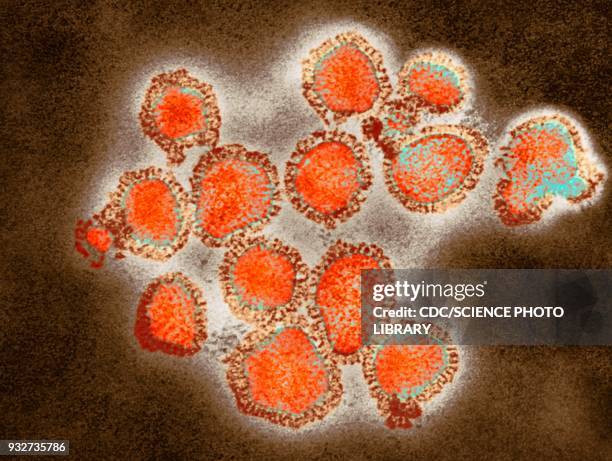
Influenza virus, Myxovirus influenzae, from the Orthomyxoviridae family. Viral diameter from 80 to 120 nanometers, taken using transmission electron...
Influenza virus, Myxovirus influenzae, from the Orthomyxoviridae family. Viral diameter from 80 to 120 nanometers, taken using transmission electron...
Influenza virus, Myxovirus influenzae, from the Orthomyxoviridae family. Viral diameter from 80 to 120 nanometers, taken using transmission electron...
Influenza virus, Myxovirus influenzae, from the Orthomyxoviridae family. Viral diameter from 80 to 120 nanometers, taken using transmission electron...
Influenza virus, Myxovirus influenzae, from the Orthomyxoviridae family. Viral diameter from 80 to 120 nanometers, taken using transmission electron...流感病毒
Influenza virus, Myxovirus influenzae, from the Orthomyxoviridae family. Viral diameter from 80 to 120 nanometers, taken using transmission electron...
Influenza virus, Myxovirus influenzae, from the Orthomyxoviridae family. Viral diameter from 80 to 120 nanometers, taken using transmission electron...
Influenza virus, Myxovirus influenzae, from the Orthomyxoviridae family. Viral diameter from 80 to 120 nanometers, taken using transmission electron...
Influenza virus, Myxovirus influenzae, from the Orthomyxoviridae family. Viral diameter from 80 to 120 nanometers, taken using transmission electron...
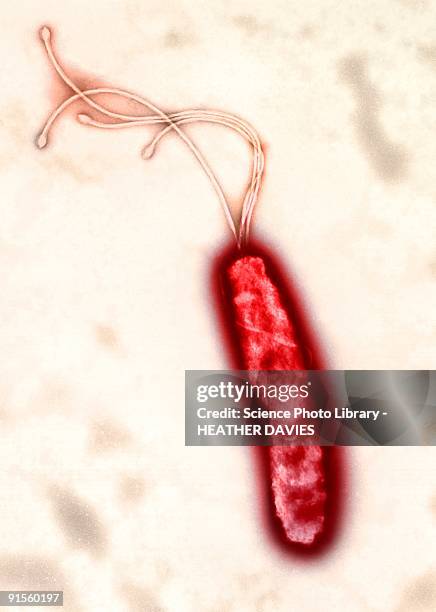
helicobacter pylori bacterium, colored transmission electron micrograph (tem) - 透射電子顯微拍攝 幅插畫檔、美工圖案、卡通及圖標科學顯微圖片, 幽門螺桿菌, 細菌
helicobacter pylori bacterium, colored transmission electron micrograph (tem) - 透射電子顯微拍攝 幅插畫檔、美工圖案、卡通及圖標幽門螺桿菌, 細菌, 顯微鏡
vaccinia virus particle (vv), colored transmission electron micrograph (tem) - 透射電子顯微拍攝 幅插畫檔、美工圖案、卡通及圖標病毒
vaccinia virus particle (vv), colored transmission electron micrograph (tem) - 透射電子顯微拍攝 幅插畫檔、美工圖案、卡通及圖標天花病毒
vaccinia virus particle (vv), colored transmission electron micrograph (tem) - 透射電子顯微拍攝 幅插畫檔、美工圖案、卡通及圖標
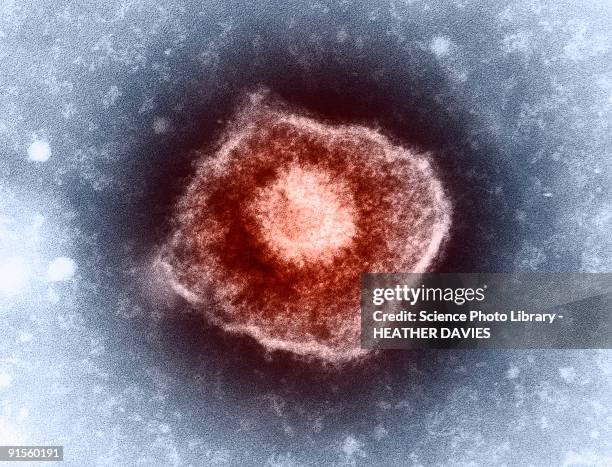
varicella zoster virus (vzv) particle, colored transmission electron micrograph (tem) - 透射電子顯微拍攝 幅插畫檔、美工圖案、卡通及圖標水痘病毒, 單純皰疹病毒