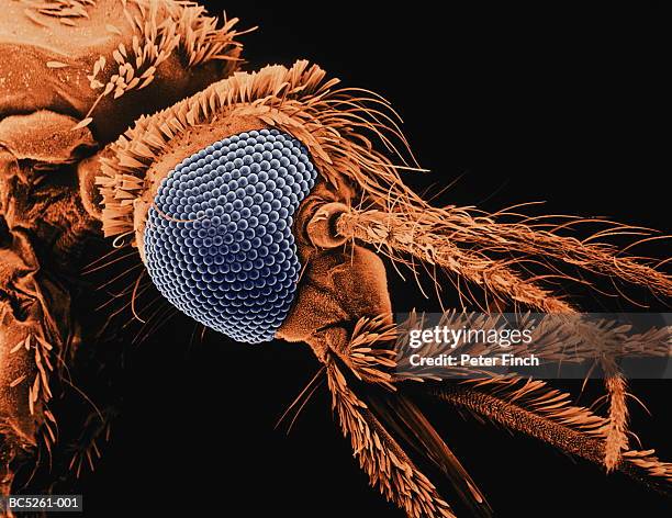
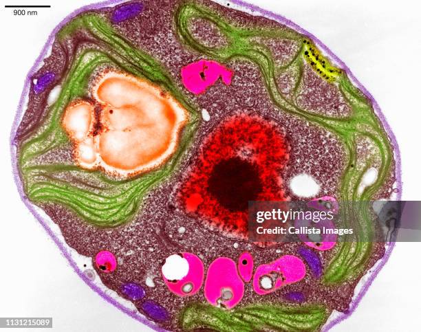
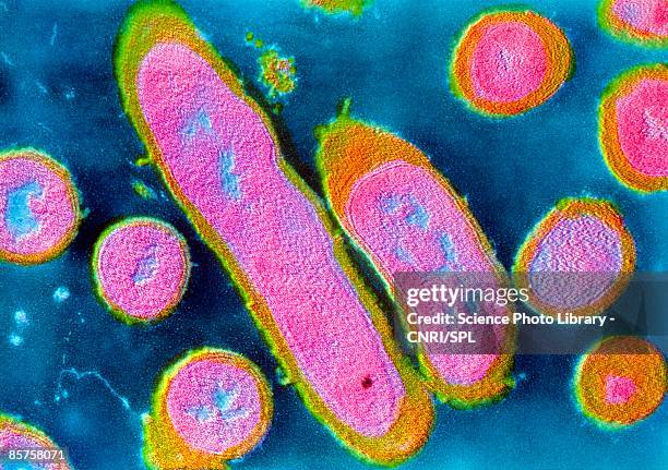
scientists using scanning electron microscope (sem) to look at pollen in biolab - 電子顯微照相 個照片及圖片檔掃描電子顯微拍攝, 電腦
vaccinia virus particle (vv), colored transmission electron micrograph (tem) - 電子顯微照相 幅插畫檔、美工圖案、卡通及圖標天花病毒
rods (pink coloured here) and cones (blue) are found in the mammalian and avian retina away from the fovea or the central zone of greatest acuity - 電子顯微照相 個照片及圖片檔細胞, 生理學, 人體構造
microscope of adenoid cystic carcinoma, rare type of cancer exist in many different body sites. - 電子顯微照相 個照片及圖片檔細胞, 抽象, 微距攝影
colorized sem of two rod-shaped carbapenem-resistant klebsiella pneumoniae (crkp) bacteria (yellow) interacting with a human neutrophil white blood cell (green) - 電子顯微照相 個照片及圖片檔克雷白氏肺炎菌
colorized sem showing numerous middle east respiratory syndrome coronavirus (mers-cov) viral particles (yellow) on the surface of a vero e6 cell (blue) - 電子顯微照相 個照片及圖片檔病毒, 掃描電子顯微拍攝
Colorized scanning electron micrograph of a cell infected with the Omicron strain of SARS-CoV-2 virus particles , isolated from a patient sample.
Colorized scanning electron micrograph of a cell infected with the Omicron strain of SARS-CoV-2 virus particles , isolated from a patient sample.
Colorized scanning electron micrograph of a cell infected with the Omicron strain of SARS-CoV-2 virus particles , isolated from a patient sample.
Textured surface details found on a honeydew melon , revealed in the 661x magnified electron micrograph film, 2004. Image courtesy Centers for...
Textured surface details found on a honeydew melon , revealed in the 2645x magnified electron micrograph film, 2004. Image courtesy Centers for...
Textured surface details found on a honeydew melon , revealed in the 661x magnified electron micrograph film, 2004. Image courtesy Centers for...
Western honeybee's head and thorax depicted in the 24x magnified scanning electron microscopic image, 2005. Image courtesy Centers for Disease...
Western honeybee's exoskeletal surface depicted in the 742x magnified scanning electron microscopic image, 2005. Image courtesy Centers for Disease...
Western honeybee's compound eye, depicted in the 2917x magnified scanning electron microscopic image, 2005. Image courtesy Centers for Disease...
Rough surface of a Vitis specimen white table grape revealed in the 5911x magnified scanning electron microscopic image, 2005. Image courtesy Centers...
varicella zoster virus (vzv) particle, colored transmission electron micrograph (tem) - 電子顯微照相 幅插畫檔、美工圖案、卡通及圖標水痘病毒, 單純皰疹病毒
colorized sem of methicillin-resistant staphylococcus aureus (mrsa) bacteria in the process of being phagocytized by a human neutrophil white blood cell - 電子顯微照相 個照片及圖片檔掃描電子顯微拍攝