human compact bone tissue,structure of bone tissue or osseous tissue, osteon - 骨髓組織 幅插畫檔、美工圖案、卡通及圖標顯微鏡, 基本組織, 矢量圖
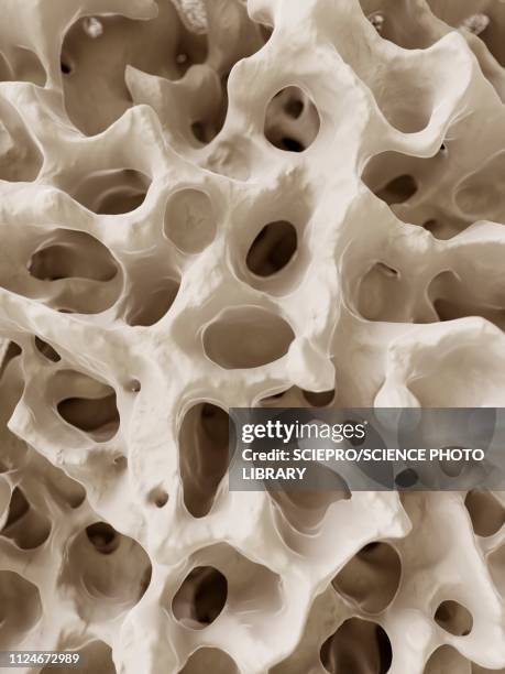
spongy (cancellous) bone. site of the red bone marrow. shows the trabeculae (bony network), red marrow fills the spaces. - 骨髓組織 個照片及圖片檔
leukemia; myeloid, 250x at 35mm. this type of leukemia has its origin in the bone marrow (myeloid tissue). it involves a malignant proliferation of immature white blood cells. this action can crowd out production of rbc s and platelets leading to an - 骨髓組織 個照片及圖片檔細胞, 癌症, 白血病
slide demonstrating breast tissue with ductal carcinoma. histopathology on the microscope. immunofluorescent photomicrograph, organs samples, histological examination, - 骨髓組織 個照片及圖片檔顯微鏡, 細胞, 抽象
compact & spongy (cancellous) bone. cross section of a long bone. shows: compact bone, spongy (cancellous) bone, and marrow cavity. - 骨髓組織 個照片及圖片檔松質骨, 人體構造
two skeleton hands on vibrant red backdrop . festive halloween background with copy space - 骨髓組織 個照片及圖片檔人類骨架, 背景 - 主題, 恐怖
antique medical scientific illustration high-resolution: nervous system - 骨髓組織 幅插畫檔、美工圖案、卡通及圖標人體, 復古風格, 人體神經系統
human bone, cross section diagram of femur showing osteon, veins, marrow. - 骨髓組織 幅插畫檔、美工圖案、卡通及圖標人體骨骼, 人體構造
Photomicrograph detecting the presence of hemosiderin, stained blue, within diffuse bone marrow, 1972. Hemosiderin, revealed here using PAS stain and...
Bone marrow smear photomicrograph of normal iron stores, prepared with Prussian blue stain, 1972. Image courtesy CDC/Dr. Gordon/D. McLaren. .
Photomicrograph of bone marrow tissue showing a normal amount of iron stores using Prussian blue staining technique, 1972. Normal iron stores are...技能
This photomicrograph of bone marrow tissue shows a normal amount of iron stores using Prussian blue stain, 1972. Normal iron stores are seen as dark...藍色
bones and osteoporosis as a medical and health concept, highlighting bone fragility, density loss, and skeletal deterioration commonly associated with aging and metabolic bone diseases. - 骨髓組織 個照片及圖片檔骨質疏鬆症, 插圖