layers of skin tissue, skin cells and fat tissue cells, dermis and adipocytes - 基本組織 幅插畫檔、美工圖案、卡通及圖標有層次
microscopic photo of a professionally prepared slide demonstrating breast tissue - 基本組織 個照片及圖片檔顯微鏡, 細胞, 分子
dense fibrous connective tissue (irregular); shows irregular collagenous fibers (pink) and fibroblast nuclei (purple); from the dermis of the skin - 基本組織 個照片及圖片檔蛋白質, 細胞, 式樣
hyaline cartilage; tracheal cartilage, supporting connective tissue, 250x at 35mm. shows: chondrocytes (cartilage cells), and matrix (intercellular material, non-living). - 基本組織 個照片及圖片檔科學顯微圖片, 細胞, 光學顯微圖
hyaline cartilage; supporting connective tissue, 250x at 35mm. shows: chondrocytes (cartilage cells), matrix (intercellular material, non-living), and lacunae (clear spaces). - 基本組織 個照片及圖片檔
motor neuron (multipolar) with cell body and many processes (mostly dendrites), spinal cord (magnification x100). this multipolar motor neuron comes from the anterior (ventral) horn of the spinal cord grey matter. the stain is methylene blue and phloxine. - 基本組織 個照片及圖片檔顯微鏡, 細胞, 人體細胞
human simple cuboidal epithelium, two kidney tubules in longitudinal section, (magnification x100) these are collecting tubules that are part of the nephron. also shows lumens and supporting blue connective tissue. mallory stain. - 基本組織 個照片及圖片檔細胞, 摄影
education anatomy and histopathology of tongue under the microscopic in laboratory. - 基本組織 個照片及圖片檔癌症, 細胞
microscopic photograph of a professionally prepared slide of liver tissue. - 基本組織 個照片及圖片檔人類臍帶, 科學顯微圖片, 人體細胞
sampling strategy of the stem tissues. immunofluorescent photomicrograph, organs samples, histological examination, histopathology on the microscope - 基本組織 個照片及圖片檔顯微鏡, 細胞, 實驗室
microscopic preparation immunofluorescent photomicrograph, organs samples, histological examination, histopathology on the microscope - 基本組織 個照片及圖片檔檢查, 細胞, 人體構造
micrograph of rat brain. science cross section. immunofluorescent photomicrograph, organs samples, histological examination, histopathology on the microscope - 基本組織 個照片及圖片檔鼠 - 動物, 抽象
pteridium aquilinum dennstaedtiaceae. immunofluorescent photomicrograph, organs samples, histological examination, histopathology on the microscope - 基本組織 個照片及圖片檔分生組織的, 顯微鏡, 細胞
slide demonstrating breast tissue with ductal carcinoma. histopathology on the microscope. immunofluorescent photomicrograph, organs samples, histological examination, - 基本組織 個照片及圖片檔顯微鏡, 細胞, 抽象
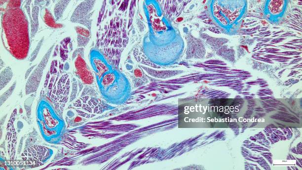
professionally prepared slide demonstrating tissue. histopathology on the microscope. immunofluorescent photomicrograph, organs samples, histological examination, - 基本組織 個照片及圖片檔癌症
normal artery in cross section (magnification x100) h & e stain. three distinct layers or tunics: tunica interna (intima), tunica media, tunica externa (adventitia). also shows endothelium and lumen with red and white blood cells. - 基本組織 個照片及圖片檔甲狀腺, 摄影
transitional epithelium of human bladder (magnification x100). h & e stain. cells near the surface are the pear-shaped basement membrane, with supporting connective tissue below - 基本組織 個照片及圖片檔上皮組織, 膀胱
Researchers works in a laboratory dedicated to grow cells to produce reconstructed human tissue at the Episkin laboratory, inside the L'oreal Center...