h&e stain, light microscopy, rheumatoid arthritis of joint showing inflammation and hyperplasia - 蘇木素伊紅染料 個照片及圖片檔類風濕關節炎, 增生, 炎症
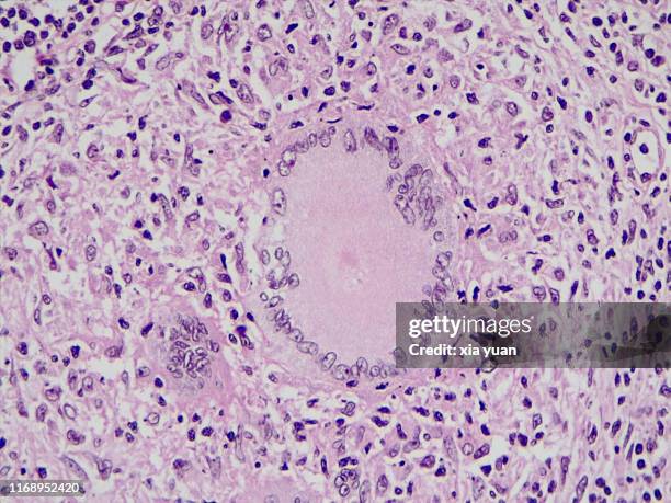
multinucleated giant cells in tuberculosis lymph nodes,40x light micrograph - 蘇木素伊紅染料 個照片及圖片檔細胞, 基本組織
h&e stain, light microscopy, healing fracture callus with exuberant cartilage and new bone formation - 蘇木素伊紅染料 個照片及圖片檔
h&e stain, light microscopy, rheumatoid arthritis of joint showing pannus - 蘇木素伊紅染料 個照片及圖片檔類風濕關節炎, 細胞
histology micrograph of renal failure,showing interstitial nephritis in tissue of kidney with h&e stain - 蘇木素伊紅染料 個照片及圖片檔
histology micrograph of renal failure,showing interstitial nephritis in tissue of kidney with h&e stain - 蘇木素伊紅染料 個照片及圖片檔
histology micrograph of renal failure,showing interstitial nephritis in tissue of kidney with h&e stain - 蘇木素伊紅染料 個照片及圖片檔
histology micrograph of renal failure,showing interstitial nephritis in tissue of kidney with h&e stain - 蘇木素伊紅染料 個照片及圖片檔炎症, 背景 - 主題, 基本組織
histology micrograph of renal failure,showing interstitial nephritis in tissue of kidney with h&e stain - 蘇木素伊紅染料 個照片及圖片檔
Photomicrograph of a monkey hair shaft processed with hematoxylin and eosin staining shows histopathologic features of white piedra due to...
skeletal muscle. longitudinal & cross sections, 250x shows: longitudinal and cross sections of skeletal muscle fibers (cells), striations, a bands (dark), i bands (light), z lines, myofibrils (longitudinal & cross sectional views), and endomysium. - 蘇木素伊紅染料 個照片及圖片檔骨骼肌
tendons have high tensile strength, light micrograph.immunofluorescent photomicrograph, organs samples, histological examination, histopathology on the microscope - 蘇木素伊紅染料 個照片及圖片檔肌肉, 顯微鏡, 細胞
human lung biopsy under microscopy. immunofluorescent photomicrograph, organs samples, histological examination, histopathology on the microscope - 蘇木素伊紅染料 個照片及圖片檔顯微鏡, 微距攝影, 細胞
lung tongue at the immunofluorescent photomicrograph, organs samples, histological examination, histopathology on the microscope - 蘇木素伊紅染料 個照片及圖片檔綠色, 生理學
immunofluorescent photomicrograph, organs samples, histological examination, histopathology on the microscope - 蘇木素伊紅染料 個照片及圖片檔脂肪肝, 顯微鏡, 細胞
lobular breast cancer, immunofluorescent photomicrograph, organs samples, histological examination, histopathology on the microscope - 蘇木素伊紅染料 個照片及圖片檔乳癌, 癌症, 細胞
micrograph of peritoneal mesothelioma. immunofluorescent photomicrograph, organs samples, histological examination, histopathology on the microscope - 蘇木素伊紅染料 個照片及圖片檔脂肪肝, 細胞, 顯微鏡
immunofluorescent photomicrograph, organs samples, histological examination, histopathology on the microscope - 蘇木素伊紅染料 個照片及圖片檔
lymph node carcinoma cells of human - immunofluorescent photomicrograph, organs samples, histological examination, histopathology on the microscope - 蘇木素伊紅染料 個照片及圖片檔
microscopic photo of a professionally prepared slide demonstrating breast tissue with ductal carcinoma. - 蘇木素伊紅染料 個照片及圖片檔乳癌, 細胞, 癌症
multinucleated giant cells in tuberculosis lymph nodes,40x light micrograph - 蘇木素伊紅染料 個照片及圖片檔顯微鏡, 彩色影像