abstract green and blue swirling line art background. a captivating abstract background featuring swirling lines in shades of green and blue, creating a mesmerizing and dynamic visual effect. - 變形蟲 個照片及圖片檔綠色, 藍色, 人體神經系統
Using trichrome staining, this photomicrograph depicts a trophozoite of the unicellular parasite, Entamoeba histolytica. Colored purple, the...
Under 500X magnification, this photomicrograph of a concentrated, iodine-stained direct mount specimen revealed the presence of a protozoan parasite...
Under 1125X magnification, this photomicrograph revealed the presence of a parasitic binucleate cyst, Entamoeba coli, which also contained a large...
This 1973 photomicrograph depicts three Naegleria gruberi, free-living amoebae. CDC/Dr George Healy 1973.
Under 1125X magnification, this photomicrograph revealed the presence of a parasitic trophozoite, Entamoeba coli. Note the single nucleus with its...
Magnified 1125X, this photomicrograph revealed the presence of a parasitic trophozoite Entamoeba histolytica, with its vacuolated cytoplasm, and...
This iodine-stained photomicrograph revealed the presence of an Entamoeba histolytica amoebic parasitic cyst. This particular organism contained...
Under 675X magnification, this photomicrograph revealed the presence of Entamoeba histolytica parasitic organisms in this ameboma tissue sample. An...
This photomicrograph revealed the presence of an Entamoeba histolytica trophozoite, which was photographed here while alive. This organism displayed...
This photomicrograph of a trichrome-stained specimen revealed the presence of an Entamoeba coli trophozoite, which contained a number of ingested...
Under 900X magnification, this photomicrograph revealed the presence of a parasitic microorganism Entamoeba coli, at the trophozoite stage, in a...
This photomicrograph of an iodine-stained specimen revealed the ultrastructural morphology of a single Entamoeba coli parasitic cyst. It appeared...
This photomicrograph was created from a cultured specimen stained with methylene blue and revealed the presence of a parasitic trophozoite, Entamoeba...
Using phase contrast imaging and magnified 1125X, this photomicrograph depicts a cyst of the protozoan Acanthamoeba polyphaga. CDC/Dr George Healy...
This scanning electron microscope image revealed some of the ultrastructural features observed on the surface of a protozoan Acanthamoeba polyphaga....
This digitally colorized scanning electron microscopic image of an untreated water specimen extracted from a wild stream mainly used to control...
Magnified 5011X, this 2002 scanning electron microscope image revealed some of the ultrastructural morphology exhibited by a small group of...
Under 500X magnification, this photomicrograph of an unstained direct mount specimen revealed the presence of a cyst of the protozoan parasite,...
set of fluid abstract blob shapes 2000s y2k style on a grid background - 變形蟲 幅插畫檔、美工圖案、卡通及圖標立體, 背景 - 主題, 插圖
abstract pink yellow neon blob quotes circle infinity amoeba shapes background - 變形蟲 個照片及圖片檔爆炸, 圓形, 無限
abstract swirl striped blob wave magical neon transparent shapes on black background. energy streams - 變形蟲 個照片及圖片檔抽象, 黑色的背景, 失真的
conceptual biomedical illustration of primary amoebic meningoencephalitis - 變形蟲 幅插畫檔、美工圖案、卡通及圖標福氏耐格里阿米巴, 傳染病, 人腦
bright poster with dynamic waves. vector illustration. abstract background. - 變形蟲 幅插畫檔、美工圖案、卡通及圖標商務, 現代 - 風格, 運動
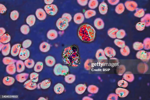
Plasmodium falciparum is a malaria-causing parasite or malaria, transmitted by the bite of female anopheles which releases merozoites into the blood...顯微鏡
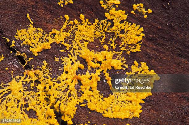
scrambled eggs slime mold. foligo septica. plasmodium. knotty stage just begun fruiting. amoeboid phase - 變形蟲 個照片及圖片檔黃色