Microphotograph of a gram stain showing flaggellated bacteria, a strain of Vibrio cholerae, the cause of Asiatic cholera, 1979. Image courtesy CDC.細菌, 腸炎弧菌
a colorized scanning electron micrograph of a white blood cell eating mrsa. - 革蘭氏染色 個照片及圖片檔革蘭氏陽性菌, 細菌
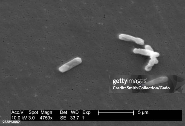
Gram-positive Clostridium difficile bacteria revealed in the stool sample micrograph film, 2004. Image courtesy Centers for Disease Control / Lois S....細菌
Gram-positive Clostridium difficile bacteria revealed in the stool sample micrograph film, 2004. Image courtesy Centers for Disease Control / Lois S....
Gram-positive Clostridium difficile bacteria revealed in the stool sample micrograph film, 2004. Image courtesy Centers for Disease Control / Lois S....
Gram-positive Clostridium difficile bacteria revealed in the stool sample micrograph film, 2004. Image courtesy Centers for Disease Control / Lois S....
Gram-positive Clostridium difficile bacteria revealed in the stool sample micrograph film, 2004. Image courtesy Centers for Disease Control / Lois S....
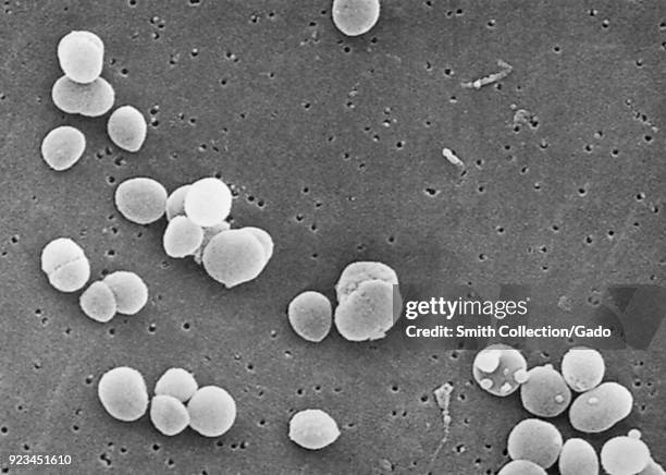
Gram-positive Staphylococcus aureus bacteria revealed in a scanning electron microscopic image, 2003. Image courtesy Centers for Disease Control .
Photomicrograph of lung tissue with various histopathologic changes indicative of Toxic Shock Syndrome , taken during an autopsy, 1984. Image...差異
Photomicrograph of Staphylococcus aureus bacteria, a causative agent of Toxic Shock Syndrome , on a Gram-stained smear, 1984. Image courtesy CDC.
colored scanning electron micrograph (sem) of many bacteria growing on blood aga - 革蘭氏染色 個照片及圖片檔掃描電子顯微拍攝, 細菌, 食物中毒
mycobacterium chelonae bacteria, colored scanning electron micrograph (sem) - 革蘭氏染色 個照片及圖片檔細菌, 掃描電子顯微拍攝, 食物中毒
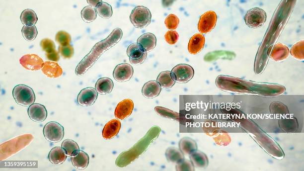
This micrograph depicts the gram-positive bacterium Propionibacterium acnes grown in thioglycollate medium at 48 hours, 1972. Propionibacterium acnes...
This micrograph depicts the gram-positive bacterium Bifidobacterium eriksonii, 1972. B. Eriksonii, also called Actinomyces eriksonii, is an anaerobic...雙歧桿菌, 細菌
This micrograph depicts histopathologic changes due to the gram-positive organism, Actinomyces israelii, 1971. Using a modified Fite-Faraco stain, a...