buttercup. ranunculus. root cross section. shows xylem, phloem, stele, starch germs & parenchyma. 100x h - 韌皮部 個照片及圖片檔
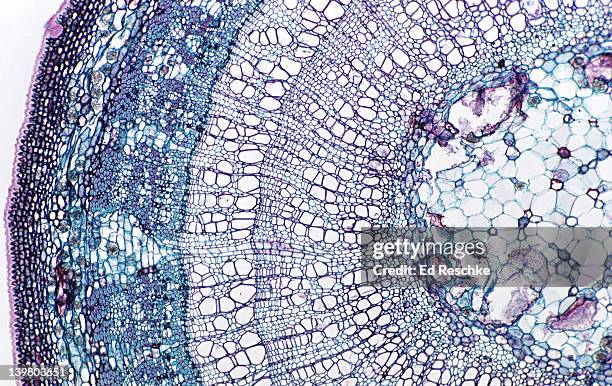
3 yr stem tilia (basswood) shows pith annual rings (3), cambium phloem, rays, periderm, cork. 10x h - 韌皮部 個照片及圖片檔
pine leaf (needle), x-section pinus sp. shows: epidermis, stoma, vascular tissue, parenchyma 25x. h - 韌皮部 個照片及圖片檔
tilia (3 year old stem). shows: cortex, xylem, phloem, pith, rays, cambium. 50x basswood.woody dicot stem h - 韌皮部 個照片及圖片檔
tilia. 3 year stem. woody dicot xylem, phloem, pith, fibers, phloem ray, annual rings (3), cambrium vascular cambium, springwood, summer wood. 5x - 韌皮部 個照片及圖片檔木髓
zea mays (indian corn). stem cross section. xylem, phloem, sclerenchyma. epidermis, parenchyma. 50x h - 韌皮部 個照片及圖片檔
x-section of buttercup mature root. ranunculus. shows stele, xylem, phloem, cortex, endodermis, starch grains & epidermis. dicot 10x - 韌皮部 個照片及圖片檔
woody dicot stem. tilia (basswood), 3 year stem, 5x. shows: 3 annual rings, xylem, phloem, rays, pith, cambium, and periderm. - 韌皮部 個照片及圖片檔
woody dicot stem. tilia (basswood), 2 year stem, 2.5x. shows: 2 annual rings, xylem, phloem, rays, pith, cambium, periderm - 韌皮部 個照片及圖片檔
woody dicot stem. tilia (basswood), 2 year stem, 25x. shows: xylem, phloem, pith, bark, cork, vascular cambium and two annual rings. - 韌皮部 個照片及圖片檔科學顯微圖片, 幹細胞, 木髓
woody dicot stem. tilia (basswood), 2 year stem, 10x. shows: 2 annual rings, xylem, phloem, rays, pith, cambium, periderm - 韌皮部 個照片及圖片檔幹細胞
woody dicot stem. tilia (basswood), 1st year stem, 10x. shows: xylem, phloem, pith, cortex, bark, cork, rays, vascular cambium, and one annual ring. - 韌皮部 個照片及圖片檔顯微鏡
stem cross section. wheat (triticum), herbaceous monocot, 50x. shows: 2 vascular bundles, xylem, phloem, epidermis, sclerenchyma, parenchyma, chlorenchyma (green areas near surface), and hollow pith. - 韌皮部 個照片及圖片檔木髓
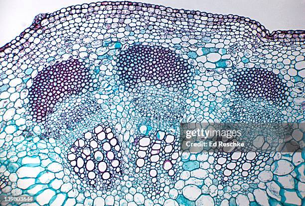
stem cross section. sunflower (helianthus), herbaceous dicot, 50x. shows a single vascular bundle. vascular bundles are arranged in a ring. shows: xylem, phloem, cambium, sclerenchyma (bundle cap), and surrounding parenchyma. - 韌皮部 個照片及圖片檔幹細胞, 背景 - 主題
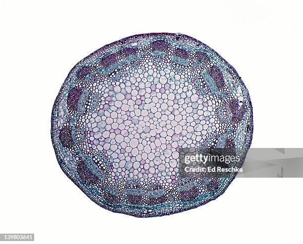
stem cross section. sunflower (helianthus), herbaceous dicot, 25x. vascular bundles arranged in a ring. shows: xylem, phloem, cambium, sclerenchyma (bundle cap), epidermis, cortex, and pith. - 韌皮部 個照片及圖片檔木髓
stem cross section. sunflower (helianthus), herbaceous dicot, 10x. shows: vascular bundles arranged in a ring, zylem, phloem, pith, cortex, and epidermis. - 韌皮部 個照片及圖片檔圓形, 顯微鏡, 幹細胞
stem cross section. sunflower (helianthus), herbaceous dicot, 8x. shows: vascular bundles arranged in a ring, xylem, phloem, pith, cortex, and epidermis. - 韌皮部 個照片及圖片檔幹細胞
stem cross section. corn (zea), herbaceous monocot, 100x. shows a single vascular bundle with: xylem, phloem, companion cells, sieve tubes, and sclerenchyma. - 韌皮部 個照片及圖片檔
stem cross section. corn (zea), herbaceous monocot, 50x. shows: scattered vascular bundles with xylem and phloem, epidermis and pith. - 韌皮部 個照片及圖片檔
stem cross section. corn (zea), herbaceous monocot, 25x. shows: scattered vascular bundles typical of monocots, xylem, phloem, sclerenchyma, pith, and epidermis. - 韌皮部 個照片及圖片檔顯微鏡, 科學顯微圖片
stem cross section. corn (zea), herbaceous monocot, 5x. shows: scattered vascular bundles typical of monocots, xylem, phloem, pith and epidermis. - 韌皮部 個照片及圖片檔幹細胞
stem cross section. buttercup (ranunculus), herbaceous dicot, 8x. shows: vascular bundles arranged in a ring (typical of dicots), xylem, phloem, epidermis, cortex, and pith. - 韌皮部 個照片及圖片檔光學顯微圖
stem cross section. basswood (tilia), 2 year stem, woody dicot, 25x. shows: 2 annual rings (springwood & summerwood), xylem, phloem rays, cortex, and cork. - 韌皮部 個照片及圖片檔顯微鏡
stem cross section. basswood (tilia), 1st year stem, woody dicot, 25x. shows: xylem, phloem, cambium, rays, pith, cork, and collenchyma. - 韌皮部 個照片及圖片檔顯微鏡
stem cross section. alfalfa (medicago), herbaceous dicot, 20x. shows: vascular bundles arranged in a ring (typical of dicots), xylem, phloem, pith, cortex, and epidermis. - 韌皮部 個照片及圖片檔特寫, 木質部, 圓形
leaf cross section, privet (ligustrum), 50x. shows: palisade mesophyll, spongy mesophyll, midvein with xylem and phloem, and epidermis. - 韌皮部 個照片及圖片檔
leaf cross section, privet (ligustrum), 50x. shows: palisade mesophyll, spongy mesophyll, stoma, epidermis, and small veins that show xylem and phloem. - 韌皮部 個照片及圖片檔
leaf cross section, privet (ligustrum), 25x. shows: palisade mesophyll, spongy mesophyll, epidermis, mid rib that contains xylem and phloem, and stoma in the lower epidermis. - 韌皮部 個照片及圖片檔
leaf cross section, privet (ligustrum), 25x. shows: palisade mesophyll, spongy mesophyll, epidermis, and a vein (a vascular bundle with xylem and phloem). - 韌皮部 個照片及圖片檔顯微鏡, 木質部, 樹葉
leaf cross section, pine (pinus), 25x. shows: photosynthetic mesophyll, xylem, phloem, endodermis, epidermis, resin ducts, and stoma. - 韌皮部 個照片及圖片檔顯微鏡
leaf cross section, lilac (syringa), 100x. shows: palisade mesophyll, spongy mesophyll, stoma, guard cells, epidermis, and a small vein that shows xylem and phloem. - 韌皮部 個照片及圖片檔顯微鏡
leaf cross section, lilac (syringa), 50x. shows: palisade mesophyll, spongy mesophyll, epidermis, stoma, guard cells, and midvein with xylem and phloem. - 韌皮部 個照片及圖片檔細胞
leaf cross section, beach grass (ammophila), sunken stomates, xerophytic adaptation. shows: photosynthetic tissue, sunken stomates, and vein (vascular bundle with xylem & phloem. 100x - 韌皮部 個照片及圖片檔植物氣孔, 下沉的
leaf cross section, beach grass (ammophila), sunken stomates, xerophytic adaptation, 50x. shows: photosynthetic tissue, sunken stomates (which represent a xerophytic adaptation), veins (vascular bundles with xylem & phloem) - 韌皮部 個照片及圖片檔顯微鏡, 摄影, 白色的背景
helianthus, sunflower stem, single vascular bundle, shows xylem, phloem, cambium, sclerenchyma 50x at 35mm - 韌皮部 個照片及圖片檔室內
microscopic view of cells of broad bean plant. immunofluorescent photomicrograph, organs samples, histological examination, histopathology on the microscope - 韌皮部 個照片及圖片檔微距攝影, 顯微鏡, 抽象
tissue in vascular plants that transports water and some nutrients. scientific research. plant tissue structure, immunofluorescent histopathology on the microscope - 韌皮部 個照片及圖片檔顯微鏡, 樹葉, 基本組織
sampling strategy of the stem tissues. immunofluorescent photomicrograph, organs samples, histological examination, histopathology on the microscope - 韌皮部 個照片及圖片檔顯微鏡, 細胞, 實驗室
dicotyledon leaf cross-section, vascular bundle, vascular tissue, immunofluorescent photomicrograph, organs samples, histological examination, histopathology on the microscope - 韌皮部 個照片及圖片檔
immunofluorescent photomicrograph, organs samples, sampling strategy of the stem tissues. histological examination, histopathology on the microscope - 韌皮部 個照片及圖片檔細胞