simple squamous epithelium (mesothelium), side or profile view, 250x. shows: the flatness of the squamous cells, nuclei, cytoplasm and cell membrane. found in the human omentum - 鱗狀上皮 個照片及圖片檔
stratified squamous epithelium (vagina, non-keratinized) 50x this epithelium has many layers (or strata) and the cells near the surface become very flat (squamous). also, shows supporting connective tissue below. - 鱗狀上皮 個照片及圖片檔
stratified squamous epithelium. vagina, non-keratinized, 100x. this epithelium has many layers (or strata) and the cells near the surface become very flat. also, shows supporting connective tissue below. - 鱗狀上皮 個照片及圖片檔
stratified squamous epithelium (vagina, non-keratinized) 100x this epithelium has many layers (or strata) and the cells near the surface become very flat (squamous). also shows supporting connective tissue below. - 鱗狀上皮 個照片及圖片檔
stratified squamous epithelium. (vagina, non-keratinized) 50x. this epithelium has many layers (or strata) and the cells near the surface become very flat (squamous). also, shows supporting connective tissue below. - 鱗狀上皮 個照片及圖片檔
stratified squamous epithelium (vagina, non-keratinized) 100x this epithelium has many layers (or strata) and the cells near the surface become very flat (squamous). also shows supporting connective tissue below. - 鱗狀上皮 個照片及圖片檔
stratified squamous epithelium. vagina, non-keratinized, 100x. this epithelium has many layers (or strata) and the cells near the surface become very flat. also, shows supporting connective tissue below. - 鱗狀上皮 個照片及圖片檔
stratified squamous epithelium (esophagus, non-keratinized, 100x) this epithelium has many layers ( or strata) and the cells near the surface become very flat (squamous). also, shows supporting connective tissue below. - 鱗狀上皮 個照片及圖片檔上皮組織, 食道
stratified squamous epithelium (esophagus, non-keratinized, 100x at 35mm) this epithelium has many layers ( or strata) and the cells near the surface become very flat (squamous). also, shows supporting connective tissue below. - 鱗狀上皮 個照片及圖片檔
photomicrograph of stratified squamous epithelium of the vagina showing desquamation of epithelial cells; note flattening of cells near surface; mallory stain, 100x. - 鱗狀上皮 個照片及圖片檔脫屑, 陰道, 上皮組織
stratified squamous epithelium epidermis (keratinized, thick skin, human, 100x). shows layers in epidermis: stratum corneum, stratum granulosum, stratum spinosum & stratum basale (basal layer of epidermis). also, shows dermis (lighter) below. - 鱗狀上皮 個照片及圖片檔有層次
stratified squamous epithelium. epidermis (keratinized, thick, human, 100x). shows layers in epidermis: stratum corneum, stratum granulosum, stratum spinosum & stratum basale (basal layer of epidermis). and dermis (lighter) below. - 鱗狀上皮 個照片及圖片檔細胞, 紫色, 特寫
normal human lung. alveoli, alveolar (air) sacs, 50x at 35mm. shows the thin alveolar walls (simple squamous epithelium) adapted for gas exchange. the red structures in the walls are red blood cells in the pulmonary capillaries. - 鱗狀上皮 個照片及圖片檔肺泡, 上皮組織, 微絲血管
alveoli. normal human lung tissue, 100x at 35mm. shows: alveoli, thin alveolar walls (simple squamous epithelium) adapted for gas exchange, red blood cells within the pulmonary capillaries, and the extremely thin respiratory membrane. - 鱗狀上皮 個照片及圖片檔微絲血管
alveoli & alveolar (air) sacs. normal lung tissue, 100x at 35mm. shows alveoli and thin alveolar walls, composed of simple squamous epithelium adapted for gas exchange. - 鱗狀上皮 個照片及圖片檔
alveolar wall, alveoli & respiratory membrane. normal human lung tissue, 250x at 35mm. shows the thin alveolar wall (simple squamous epithelium) and the extremely thin respiratory membrane beautifully adapted for gas exchange. also, shows red blood - 鱗狀上皮 個照片及圖片檔
alveolar (air) sac, alveoli, normal lung tissue, 50x at 35mm. shows a single large alveolar (or air) sac and many alveoli. the alveolar walls are composed of simple squamous epithelium. - 鱗狀上皮 個照片及圖片檔肺泡, 上皮組織
human simple cuboidal epithelium, kidney tubules, cross and longitudinal sections, (magnification x100) shows simple cuboidal epithelium forming kidney tubules (part of the nephron), lumen and blue connective tissue. a few of the tubules show flatter (squ - 鱗狀上皮 個照片及圖片檔
stratified squamous epithelium skin, epidermis, cornified (dead) layer, dermis (blue) 250x - 鱗狀上皮 個照片及圖片檔顯微鏡, 細胞, 死去
cervix. stratified squamous epithelium, microscopic anatomy (or histology), human, 150x - 鱗狀上皮 個照片及圖片檔
vector illustration of kids alphabet coloring book page with outlined clip art to color. letter c for cuboid. - 鱗狀上皮 幅插畫檔、美工圖案、卡通及圖標幾何圖形, 教師
taste bud--contains taste receptors (sensory receptors or chemoreceptors), tongue, 100x - 鱗狀上皮 個照片及圖片檔味蕾, 科學顯微圖片
simple squamous epithelium (mesothelium) forming part of the peritoneum--a serous membrane that lines the abdominal cavity, 250x - 鱗狀上皮 個照片及圖片檔
taste buds--contain taste receptors--sensory receptors or chemoreceptors, tongue, 100x - 鱗狀上皮 個照片及圖片檔
artery anatomy--shows layers or tunics, tunica interna (intima), tunica media and tunica externa (adventitia), 100x - 鱗狀上皮 個照片及圖片檔
human lung biopsy under microscopy. immunofluorescent photomicrograph, organs samples, histological examination, histopathology on the microscope - 鱗狀上皮 個照片及圖片檔顯微鏡, 微距攝影, 細胞
lung tongue at the immunofluorescent photomicrograph, organs samples, histological examination, histopathology on the microscope - 鱗狀上皮 個照片及圖片檔綠色, 生理學
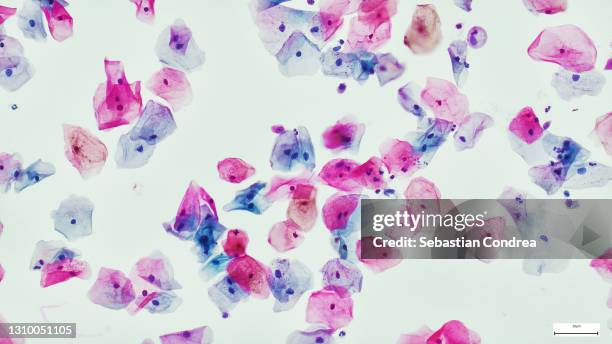
squamous epithelial cells of human cervix under the microscope view. pap smear test is a procedure to test for cervical cancer in women - 鱗狀上皮 個照片及圖片檔人類乳突病毒, 細胞, 顯微鏡
lymph node carcinoma cells of human - immunofluorescent photomicrograph, organs samples, histological examination, histopathology on the microscope - 鱗狀上皮 個照片及圖片檔
human liver tissue under the microscope, immunofluorescent photomicrograph, organs samples, histological examination, histopathology on the microscope - 鱗狀上皮 個照片及圖片檔
taste buds--contain taste receptors (sensory receptors or chemoreceptors), tongue, 100x - 鱗狀上皮 個照片及圖片檔複層鱗狀上皮
anatomy of an artery--tunica interna (or intima), tunica media, tunica externa (or adventitia), 100x - 鱗狀上皮 個照片及圖片檔
stratified squamous epithelium of human vagina (magnification x 50) h & e stain. non-keratinized. cells become very flat near surface. - 鱗狀上皮 個照片及圖片檔陰道, 基本組織
skin; scalp, human, capillaries in dermis, 100x at 35mm. shows: epidermis (stratified squamous epithelium, keratinized), dermis with capillaries and red blood cells. - 鱗狀上皮 個照片及圖片檔
skin. epidermis, dermis, subcutaneous layer, human, thick skin, 25x at 35mm. shows: epidermis (keratinized stratified squamous epithelium), dermis, subcutaneous layer, sweat glands and duct, adipose tissue. - 鱗狀上皮 個照片及圖片檔光學顯微圖
meissners corpuscles. sensory receptors (tactile) in thick skin from finger, 50x at 35mm. these are tactile (touch) sensory receptors in the dermis. shows layers of the epidermis (stratified squamous epithelium), the dermis & meissners corpuscles. - 鱗狀上皮 個照片及圖片檔